| Parasite | Loma sp. (Loma salmonae ?) |
|---|---|
| Taxonomy | Microspora, Microsporea |
| Host | Red spotted masu salmon (Oncorhyncus masou) |
| Disease name | Microsporidial gill disease |
| Infection site | Gill |
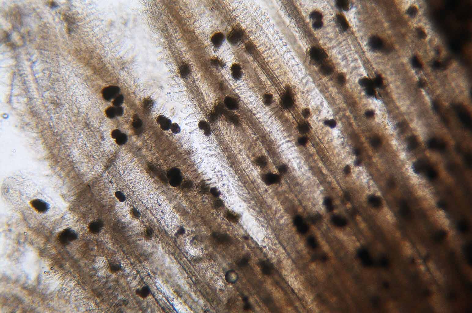
| Clinical signs | Many spherical xenomas (a complex formed by a host cell and a parasite) are observed in the gill (Figs. 1, 2). |
| Parasitology | A number of spores are produced inside the xenomas (Fig. 3). A spore (average length 4.5 mm; average width 2.3 mm) is pyriform. |
| Pathology | The gill filaments were swollen and clubbing. |
| Health hazard | Since this parasite is not infectious to human, it is harmless in food hygiene. |
| Diagnosis | Check the spores by the wet-mount of xenomas. The sample should be smeared and stained by Uvitex 2B followed by a fluorescent microscopic observation. The stained spores emit blue fluorescence under UV radiation. |
| Other information | Loma salmonae is well known species because it causes heavy damage to the culture of Chinook salmon Oncorhynchus tshawytscha and coho salmon O. kisutch in North America. In Japan, Loma salmonae-like species was reported in masu salmon O. masou, but this microsporidium is not identified to the species level. (Awakura et al., 1982). Likewise, Loma sp. in red spotted masu salmon is not identified as Loma salmonae despite its morphological similarities. |
| References | Awakura, T., M. Tanaka and M. Yoshimizu (1982):
Studies on parasites of masu salmon, Oncorhynchus
masou- IV Loma sp. (Protozoa:
Microsporea) found in the gills. Sci. Rep. Hokkaido Fish Hatchery, 37, 49-55. |
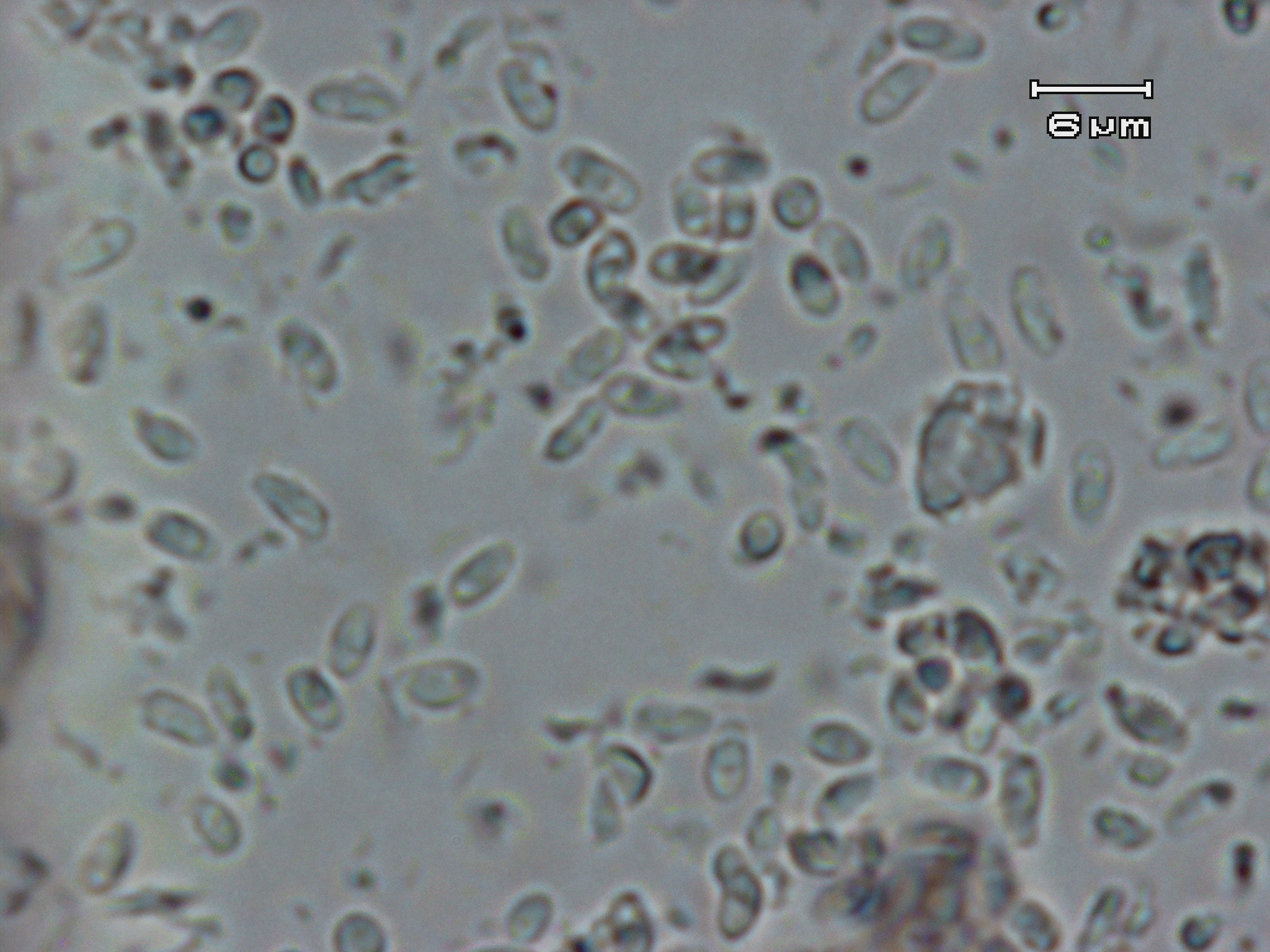
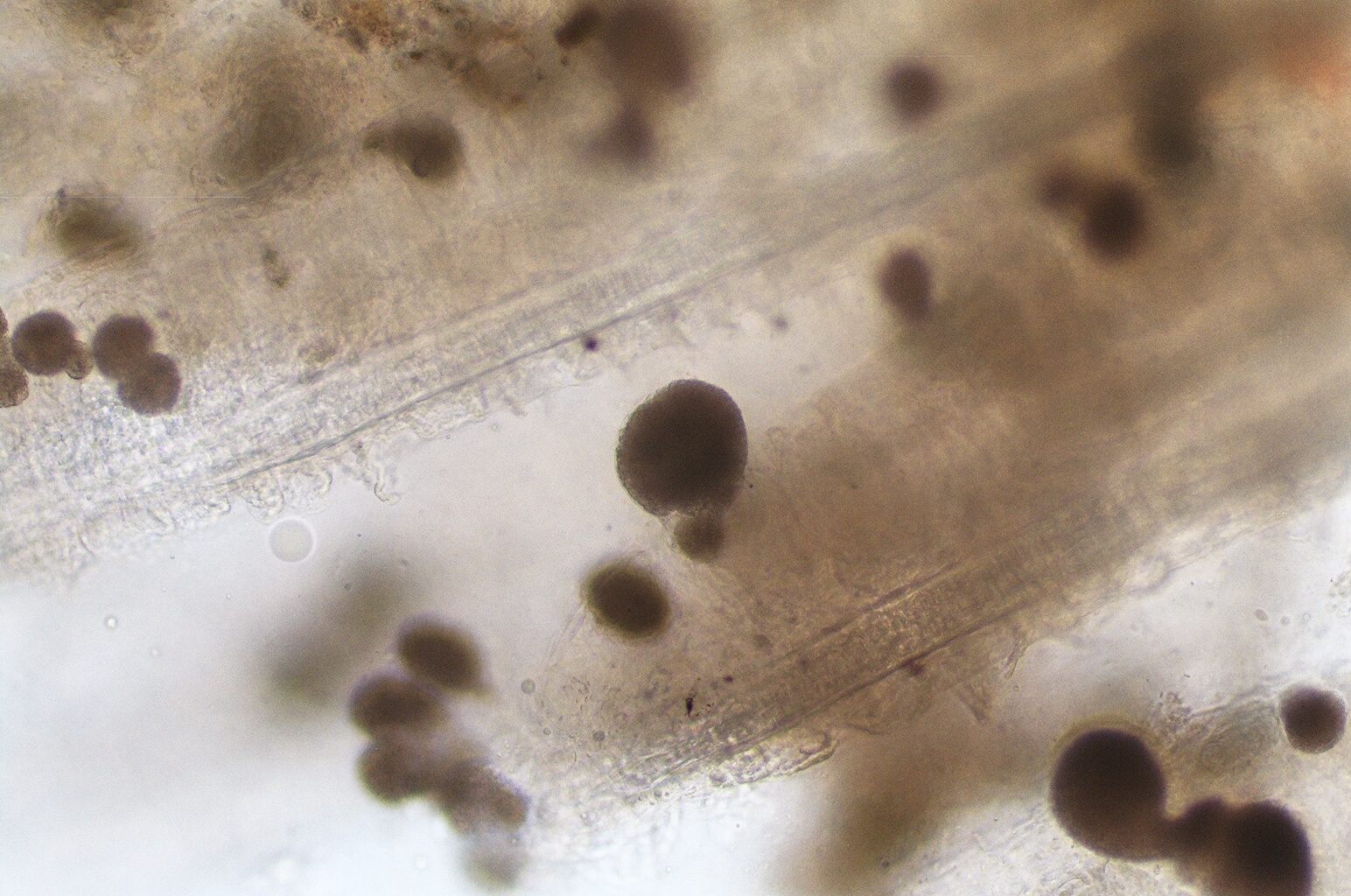

Fig. 3. Fresh spores of Loma sp.
(Photos by H. Aoshima (1, 2))
Fig. 1. Numerous xenomas formed in the gill filament.
Fig. 2. Xenomas of Loma sp.